Mouse Embryo
Magnetic Resonance Imaging
Mouse Embryo
Magnetic Resonance Imaging
The Mouse Embryo Magnetic Resonance Imaging website is a collection of images depicting the development of the mouse from days 9.5 to 19.5 days after fertilization. The site presents magnetic resonance (MR) images for each stage of development, including image slices from three distinct views (sagittal, coronal, and axial), whole embryo views, and animations. The site includes a page with several examples of magnetic resonance angiography (MRA).
The collection of images is intended to serve students, researchers, and the general public interested in viewing, studying and teaching animal development. The site also serves as an historical record of the early state of MR imaging of small specimens.

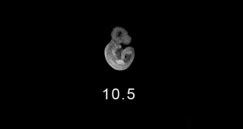
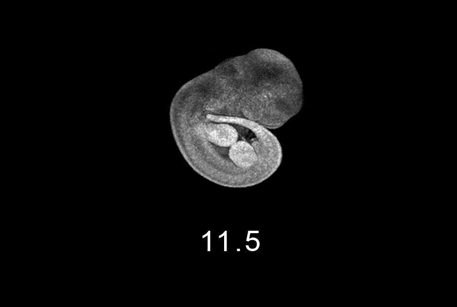
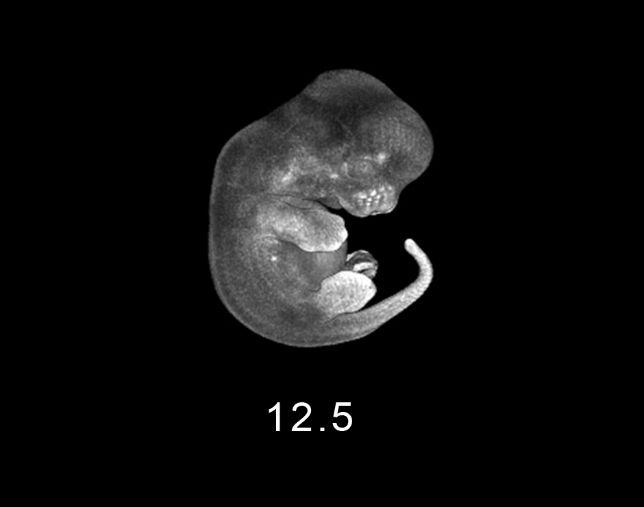
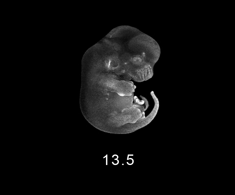
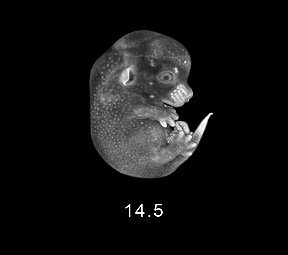
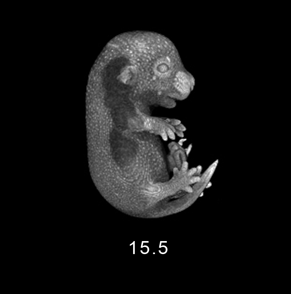
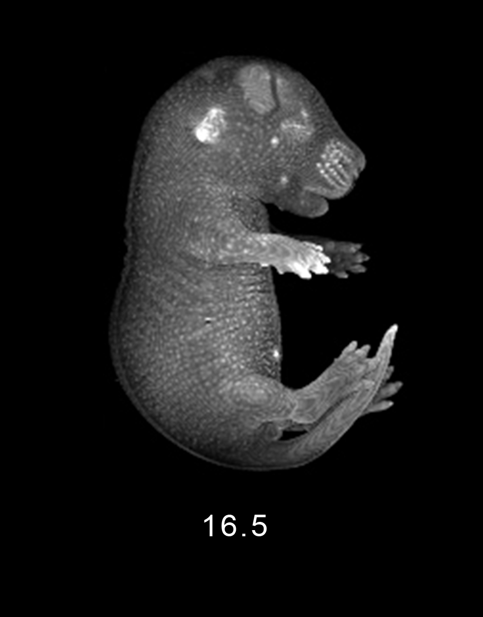
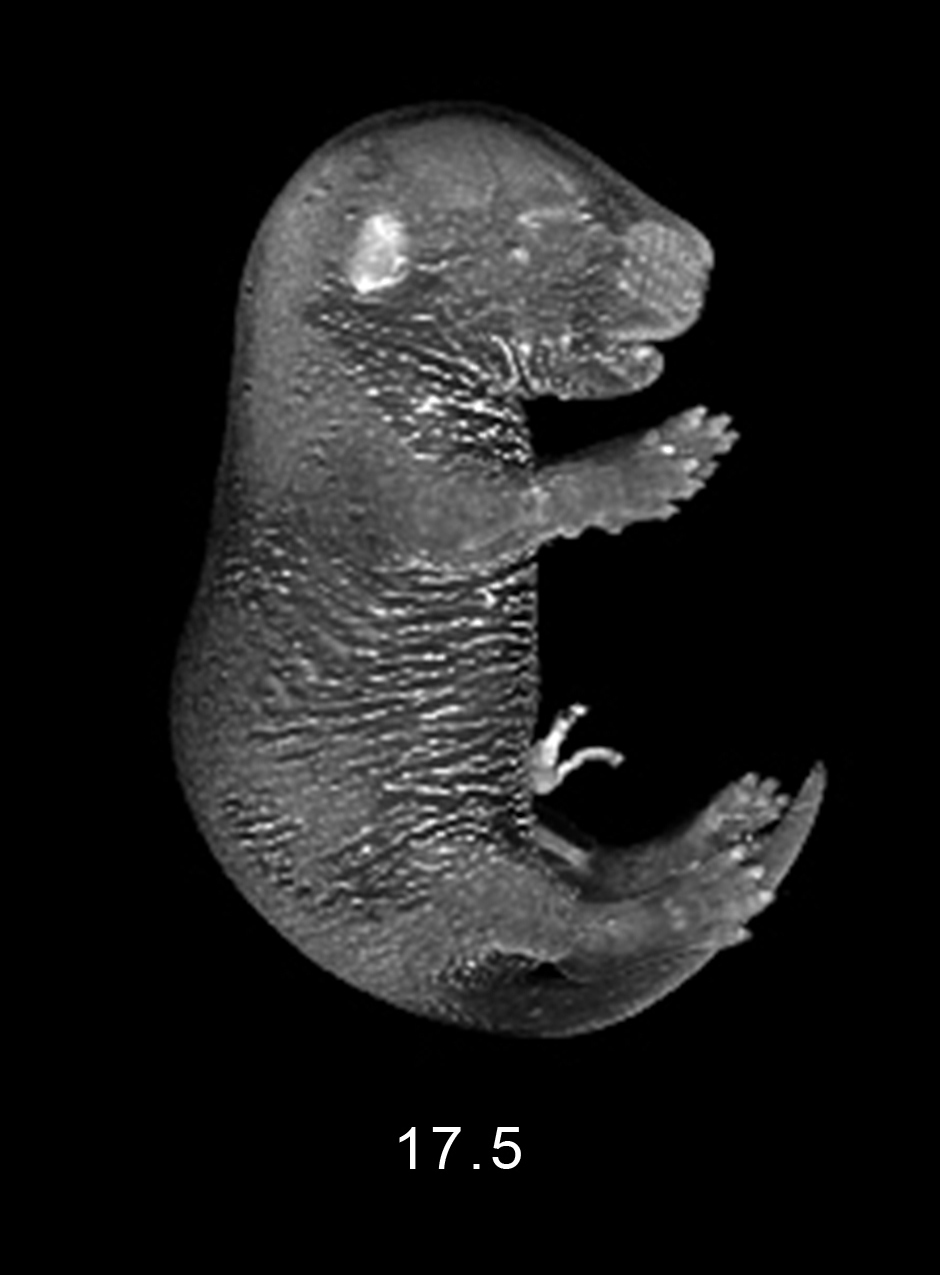

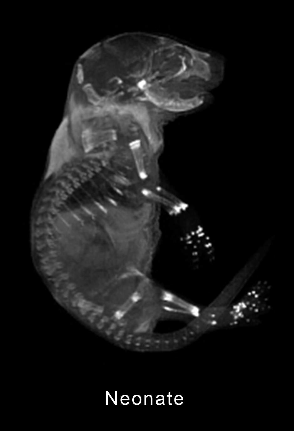

The age of a mouse embryo is typically presented in gestational days or as the number of days post-fertilization (days post coitum, or dpc). The half-day demarcation is due to the typical breeding procedure with mating set up in the late afternoon, with noontime after a vaginal plug is detected in the morning, being defined as 0.5 dpc. The mouse is typically born about day 19 post-fertilization.
Original imaging was performed by Brad Smith in the mid-1990's at the Center for In-vivo Microscopy (CIVM) at Duke University with valuable support from Elwood Linney (mouse development) and Mark Johnson (MR microscopy). The CIVM was an early leader in magnetic resonance microscopy and MR imaging of small animals. Later image processing and the production of animations was performed by Brad while at the School of Art & Design, University of Michigan.
© 2026.
Mouse Embryo Magnetic Resonance Imaging by Brad Smith is licensed under CC BY-NC-SA 4.0![]()
![]()
![]()
![]()