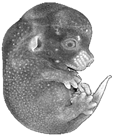
Mouse Embryo
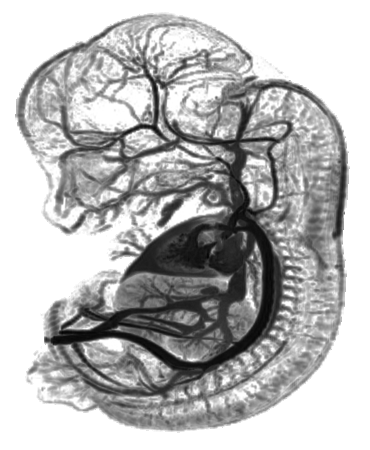
Magnetic Resonance Imaging
Mouse Embryo
Magnetic Resonance Imaging
Methods
Magnetic resonance imaging (MRI) and magnetic resonance angiography (MRA) of mouse embryos were performed according to methods developed by Brad Smith and colleagues* and included adaptations to proceedures developed and reported for mouse microangiography by Eric L. Effmann**.
Preparation and Perfusion of Embryos
Mice were maintained according to institutional guidelines. Embryos were surgically extracted from the anesthetized female mouse (2.5% Avertin , .015ml/gram body weight). Phosphate-buffered saline was perfused into the umbilical vein of the embryo followed by a fixative perfusion (2% glutaraldehyde and 1% formaline in 300-mOsm/L phosphate buffer). For magnetic resonance angiography, an MRI contrast agent, dissolved in a 10% gelatin solution, was perfused through the umbilical artery. After the perfusions, the embryos were quickly immersed in cold (4ºC) phosphate-buffered saline to solidify the gelatin and then immersion fixed.
Perfusion was performed through a four-channel Gilson peristaltic pump and finely drawn glass micropipette (25-75 micrometer tip diameter) at flow rates of approximately 2-3 microliters per minute. A small tear in the umbilical artery was created to receive the perfusion micropipette and an additional small tear in the umbillical vein served to vent fluid as the perfusion fluids were introduced. The umbilical vessels were ligated prior to the embryo being immersion fixed in the perfusion fixative and until they were embedded in 3% low melting-point agarose for MR microscopy analysis.
Magnetic Resonance Imaging
Magnetic resonance imaging was performed at the Center for In-vivo Microscopy (CIVM) at Duke University. MRI data were acquired at 9.4 T using a GE NMR Instruments Omega system modified for MR microscopy using a custom-built 1 cm solenoid rf coil constructed from a sheet of polyfon microwave substrate. Data were acquired using three-dimensional spin warp encoding with TR = 200 ms, TE = 6 ms, and four excitations for each phase-encoding step. Imaging data were reconstructed resulting in 256 isotropic (equal x, y, z resolution), 16-bit image slices.
Volume rendering and animations were produced using VoxelView Ultra 2.0 software (Vital Images, Fairfield, IO).
Citations
* Smith BR, Johnson GA, Groman EV, Linney E. Magnetic Resonance Microscopy of Mouse Embryos. Proceedings of the National Acadmemy of Science, USA 1994; 91:3530-3533.
* Smith BR. Magnetic Resonance Microscopy in Cardiac Development. Microscopic Research Techniques 2001; 52:323-330.
** Effmann EL, Whitman S, Pexieder T. Stereo microangiography in embryologic and teratologic investigation. Teratology. 1986 Aug;34(1):103-12.
© 2026.
Chick Embryo Microangiography by Brad Smith is licensed under CC BY-NC-SA 4.0![]()
![]()
![]()
![]()